

このスライドは会員限定コンテンツです。
今すぐアカウント登録(無料)して、続きを読みましょう!


1/44
関連するスライド

中枢神経感染症
森本将矢
27547
125
"代替薬時代"の感染症外来
高野哲史
92538
211

抗菌薬+αのコントロバシー
藤原翔
73526
134
β-ラクタム系抗菌薬 -カルバペネム系とその他のβ-ラクタム系薬-
高野哲史
29724
134
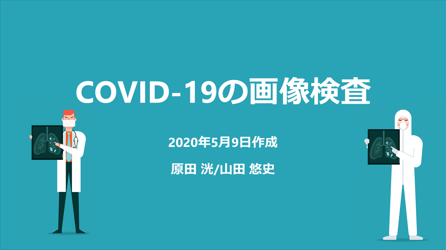
COVID-19の画像検査
The Mount Sinai Hospital
74,162
109
概要
COVID-19の画像検査についてのスライドです。下記のクリニカルクエスチョンについてエビデンスベースでまとめています。【胸部X線は診断に有効か?】【
胸部CTの感度・特異度は?】【胸部CTの特徴的な所見は?】【重症化するCOVID-19の胸部CTの特徴は?】【無症候の患者に胸部CTの異常はみられるか?】岡山大学病院内科専攻医 原田 洸先生と協力して作成しました。(2020年5月9日作成)
【このスライドの解説動画はこちら】
本スライドの対象者
研修医
投稿された先生へ質問や勉強になったポイントをコメントしてみましょう!
0 件のコメント





山田悠史さんの他の投稿スライド
すべて見る

このスライドと同じ診療科のスライド


テキスト全文
COVID-19の画像検査の概要と症例提示
#1.
2020年5月9日作成 原田 洸/山田 悠史 COVID-19の画像検査
#2.
症例提示:70歳男性 発熱、咳嗽 5日前から続く発熱、咳嗽、呼吸困難あり。近医より、COVID-19の疑いで紹介となった。 問診・身体診察の後、画像検査を行うことに。 もしもCOVID-19であれば、どのような所見がみられるだろうか? ※架空の症例です
胸部X線とCTの診断有効性
#3.
クリニカルクエスチョン 胸部X線は診断に有効か? 胸部CTの感度・特異度は? 胸部CTの特徴的な所見は? 重症化するCOVID-19の胸部CTの特徴は? 無症候の患者に胸部CTの異常はみられるか?
胸部X線の異常所見とその分布
胸部X線の診断結果のまとめ
胸部CTの感度・特異度の評価
胸部CTの特徴的な所見と病変の分析
重症化するCOVID-19の胸部CTの特徴
無症候患者におけるCTの異常所見
症例の転帰とCOVID-19画像所見のまとめ
#4.
基礎知識:GGOとconsolidation すりガラス影 (GGO: Ground Glass Opacity)肺血管の辺縁が認知できる程度の肺野の吸収値の増大。 浸潤影 (Consolidation)容積減少を伴わず、肺血管の辺縁を覆い隠す肺野の吸収値の増大で、肺血管が埋もれて見えない状態。 Radiological findings from 81 patients with COVID-19 pneumonia in Wuhan, China: a descriptive studyhttps://www.thelancet.com/article/S1473-3099(20)30086-4/
#5.
胸部X線は診断に有効か?
#6.
胸部X線は有効? Frequency and Distribution of Chest Radiographic Findings in COVID-19 Positive Patientshttps://pubs.rsna.org/doi/10.1148/radiol.2020201160
#7.
胸部X線は有効? 香港でCOVID-19が記録された64人の患者を対象とした後方視的研究。 69%が入院時に胸部X線に異常があり、80%が入院中にX線に異常があった。 20%の患者が発病中のどの時点でも胸部X線に異常を認めなかった。 Frequency and Distribution of Chest Radiographic Findings in COVID-19 Positive Patientshttps://pubs.rsna.org/doi/10.1148/radiol.2020201160
#8.
GGOやconsolidationがみられる Frequency and Distribution of Chest Radiographic Findings in COVID-19 Positive Patientshttps://pubs.rsna.org/doi/10.1148/radiol.2020201160
#9.
胸部X線ではわかりにくい場合もある Frequency and Distribution of Chest Radiographic Findings in COVID-19 Positive Patientshttps://pubs.rsna.org/doi/10.1148/radiol.2020201160
#10.
胸部X線は有効? 胸部X線の異常所見はGGOやconsolidationであり、両側性、末梢性、下葉に分布していた。 肺病変は発病の経過とともに増加し発症後10~12日目に胸部X線所見の重症度のピークを迎えた。 Frequency and Distribution of Chest Radiographic Findings in COVID-19 Positive Patientshttps://pubs.rsna.org/doi/10.1148/radiol.2020201160
#11.
まとめ:胸部X線は診断に有効か? ・80%の患者で、発症後10~12日をピークに、 入院中のどこかで胸部X線の異常がみられる。 ・単純X線では初期には異常がとらえにくい。 ・胸部画像が必要な症例では、感染予防策のもとで 胸部CTを検討する。
#12.
症例の続き ・70歳男性の発熱・咳嗽。 ・胸部X線では両側肺野に GGOを認めた。 ・胸部CTではどのような所見がみられるか?
#13.
胸部CTの感度・特異度は?
#14.
胸部CTの感度・特異度は? Correlation of Chest CT and RT-PCR Testing in Coronavirus Disease 2019 (COVID-19) in China: A Report of 1014 Caseshttps://pubs.rsna.org/doi/10.1148/radiol.2020200642
#15.
胸部CTの感度・特異度は? 2020年1月6日~2月6日に武漢市でCOVID-19が疑われた2~95歳の1,014名(99%は成人)を対象に、胸部CTとRT-PCR検査の両方を受けた患者を評価した。 検査の陽性率は、RT-PCR: 59% (601/1014) < 特徴的なCT所見: 88% (888/1014) ※特徴的CT所見: 両側GGOを主体としたウイルス肺炎像 Correlation of Chest CT and RT-PCR Testing in Coronavirus Disease 2019 (COVID-19) in China: A Report of 1014 Caseshttps://pubs.rsna.org/doi/10.1148/radiol.2020200642
#16.
胸部CTの感度・特異度は? RT-PCRをreferenceとすると、胸部CTの診断性能は感度: 97%、特異度: 25% 60~93%の患者で、 RT-PCR検査が陽性になる前にCOVID-19に合致するCT所見がみられた。 42%の患者でRT-PCRの結果が陰性になる前に、胸部CTフォローの所見で改善を認めた。 Correlation of Chest CT and RT-PCR Testing in Coronavirus Disease 2019 (COVID-19) in China: A Report of 1014 Caseshttps://pubs.rsna.org/doi/10.1148/radiol.2020200642
#17.
まとめ:胸部CTの感度・特異度は? 胸部CTは、PCRを基準にすると感度: 97%、特異度: 25%。 6~9割の患者で、PCRが陽性になる前にCT所見の異常がみられるかもしれない。
#18.
胸部CTの特徴的な所見は?
#19.
メタアナリシス CT Imaging Features of 4121 Patients With COVID-19: A Meta-Analysis https://onlinelibrary.wiley.com/doi/full/10.1002/jmv.25910
#20.
メタアナリシス COVID-19患者 4121人のCT所見を評価した34件の後方視研究のシステマティックレビュー。 2020年1月1日~3月16日に発表された論文が対象。 胸部CTでの病変の分布、病変の密度や性状、形態、随伴所見などを解析した。 CT Imaging Features of 4121 Patients With COVID-19: A Meta-Analysis https://onlinelibrary.wiley.com/doi/full/10.1002/jmv.25910
#21.
特徴的なCT所見:分布 両側 多葉 単一 単葉 正常 74% 68% 19% 15% 8% [%] CT Imaging Features of 4121 Patients With COVID-19: A Meta-Analysis https://onlinelibrary.wiley.com/doi/full/10.1002/jmv.25910
#22.
特徴的なCT所見:病変の密度 GGO Air bronchogram Crazy paving pattern Consolidation [%] CT Imaging Features of 4121 Patients With COVID-19: A Meta-Analysis https://onlinelibrary.wiley.com/doi/full/10.1002/jmv.25910
#23.
特徴的なCT所見:病変の密度 Radiological findings from 81 patients with COVID-19 pneumonia in Wuhan, China: a descriptive studyhttps://www.thelancet.com/article/S1473-3099(20)30086-4/ ・Air bronchogram ・Crazy paving pattern GGOの内部に網状影を伴う所見 気管支周囲の肺胞腔の含気が消失し気管支が透亮して見える
#24.
特徴的なCT所見:病変の形 Patchy lesion Spider web sign 線状陰影 結節影 [%] CT Imaging Features of 4121 Patients With COVID-19: A Meta-Analysis https://onlinelibrary.wiley.com/doi/full/10.1002/jmv.25910
#25.
特徴的なCT所見:病変の形 Chest CT Findings in Patients With Coronavirus Disease 2019 and Its Relationship With Clinical Featureshttps://www.ncbi.nlm.nih.gov/pmc/articles/PMC7147284/ ・Patchy lesion ・Spider web sign 隣接する胸膜が引っ張られクモの巣のような形にみえる陰影 斑状の陰影 CT features of SARS-CoV-2 pneumonia according to clinical presentation: a retrospective analysis of 120 consecutive patients from Wuhan cityhttps://link.springer.com/article/10.1007%2Fs00330-020-06854-1
#26.
特徴的なCT所見:病変の形 ・線状陰影 (Cord-like pattern) 過去の炎症の痕跡としてみられる線維化 ・結節影 (Nodular) lesion) Imaging characteristics of initial chest computed tomography and clinical manifestations of patients with COVID-19 pneumoniahttps://www.ncbi.nlm.nih.gov/pmc/articles/PMC7171599/ COVID-19では結節状のGGOがみられることもある
#27.
特徴的なCT所見:随伴所見 胸膜肥厚 リンパ節腫脹 胸水 5% [%] CT Imaging Features of 4121 Patients With COVID-19: A Meta-Analysis https://onlinelibrary.wiley.com/doi/full/10.1002/jmv.25910
#28.
まとめ:胸部CTの特徴的な所見は? ・COVID-19 では「両側・多葉に多発するGGO」が 典型的な所見であるが、他のウイルス性肺炎などでも みられるため特異的とは言えない。 ・リンパ節腫脹や胸水を伴うことは珍しい。
#29.
重症化するCOVID-19の胸部CTの特徴は?
#30.
重症例の胸部CTの特徴は? CT features of SARS-CoV-2 pneumonia according to clinical presentation: a retrospective analysis of 120 consecutive patients from Wuhan cityhttps://link.springer.com/article/10.1007%2Fs00330-020-06854-1
#31.
重症例の胸部CTの特徴は? ・2020年1月10日~2月10日に武漢の病院(単施設)に 入院したCOVID-19肺炎患者120例を評価した。 ・無症状: 16名、軽度・中等度:74 名、重症: 30名であった。 ・無症状~中等症の90名と重症30名のCT所見を比較した。 CT features of SARS-CoV-2 pneumonia according to clinical presentation: a retrospective analysis of 120 consecutive patients from Wuhan cityhttps://link.springer.com/article/10.1007%2Fs00330-020-06854-1
#32.
非重症 vs 重症 両側病変 GGO Crazy paving pattern Consolidation 結節 線状の病変 [%] 赤字: p<0.05 CT features of SARS-CoV-2 pneumonia according to clinical presentation: a retrospective analysis of 120 consecutive patients from Wuhan cityhttps://link.springer.com/article/10.1007%2Fs00330-020-06854-1
#33.
非重症 vs 重症 気管支拡張 胸水 Tree-in-bud sign 全葉の病変 Air bronchogram White lung* [%] リンパ節腫脹 赤字: p<0.05 *White lung =完全に片肺真っ白 CT features of SARS-CoV-2 pneumonia according to clinical presentation: a retrospective analysis of 120 consecutive patients from Wuhan cityhttps://link.springer.com/article/10.1007%2Fs00330-020-06854-1
#34.
非重症 vs 重症 ・Consolidation、Air bronchogram、White lung、胸水、 Crazy paving pattern、線状病変、気管支拡張などが 重症患者で多くみられた。 ・多変量解析の結果、 Crazy paving pattern (OR = 15.3, 95% CI = 2.6-89.5) や Air bronchogram (OR = 41.8, 95% CI = 5.9–298.4) は 重症化リスクと統計的に有意な相関がみられた。 CT features of SARS-CoV-2 pneumonia according to clinical presentation: a retrospective analysis of 120 consecutive patients from Wuhan cityhttps://link.springer.com/article/10.1007%2Fs00330-020-06854-1
#35.
まとめ:重症化するCOVID-19の胸部CTの特徴は? ・重症化するCOVID-19では、Consolidationや Air bronchogram、Crazy paving patternなどが 非重症例よりも多くみられる。
#36.
無症候の患者に胸部CTの異常はみられるか?
#37.
Chest CT Findings in Cases from the Cruise Ship “Diamond Princess” with Coronavirus Disease 2019 (COVID-19)https://pubs.rsna.org/doi/10.1148/ryct.2020200110 無症候患者の胸部CTの異常は?
#38.
・単施設(自衛隊中央病院)の後方視的研究。 ・ダイヤモンド・プリンセス号の乗員乗客で、 PCR で COVID-19 が確認された 104 例(平均年齢 62 歳) を対象とし、CT画像を評価した。 ・全体の73%が無症状、27%が有症状であった。 無症候患者の胸部CTの異常は? Chest CT Findings in Cases from the Cruise Ship “Diamond Princess” with Coronavirus Disease 2019 (COVID-19)https://pubs.rsna.org/doi/10.1148/ryct.2020200110
#39.
・76人の無症候患者のうち、54%にあたる41人で CTでの異常所見を認めた。 ・無症候患者のCT所見のうち 83%はGGO主体、 17%はconsolidation主体。 無症候患者の胸部CTの異常は? GGO 83% Consolidation17% Chest CT Findings in Cases from the Cruise Ship “Diamond Princess” with Coronavirus Disease 2019 (COVID-19)https://pubs.rsna.org/doi/10.1148/ryct.2020200110
#40.
無症状患者のCT所見の例 ・70歳女性 無症状・両肺野にGGOあり・air bronchogramを伴う ・73歳女性 無症状・両側下肺野に気管支拡張あり・GGOとconsolidationあり Chest CT Findings in Cases from the Cruise Ship “Diamond Princess” with Coronavirus Disease 2019 (COVID-19)https://pubs.rsna.org/doi/10.1148/ryct.2020200110
#41.
まとめ:無症候の患者に 胸部CTの異常はみられるか? ・無症候PCR陽性患者の約半数で、CT所見の 異常を認める可能性がある。 ・無症候の場合、CT所見はGGO>consolidationの パターンが多い。
#42.
症例の転帰 ・70歳男性の発熱・咳嗽。 ・CTで両側肺野に多発するGGOを認め、 COVID-19を疑う所見であった。 ・SARS-CoV2の鼻咽頭のPCR検査を行い、陽性であった。
#43.
COVID-19 画像所見のまとめ ・単純X線では初期には異常がとらえにくい。 ・CTでは「両側・多葉に多発するGGO」が 典型的な所見である。 ・無症候患者でもGGO主体の異常がみられることがあり、 重症例ではConsolidation、Air bronchogram、 Crazy paving pattern などが比較的多くみられる。
会員登録・ログインで「スライド内のテキスト」を参照いただけます。