テキスト全文
グラム染色の重要性と実施方法
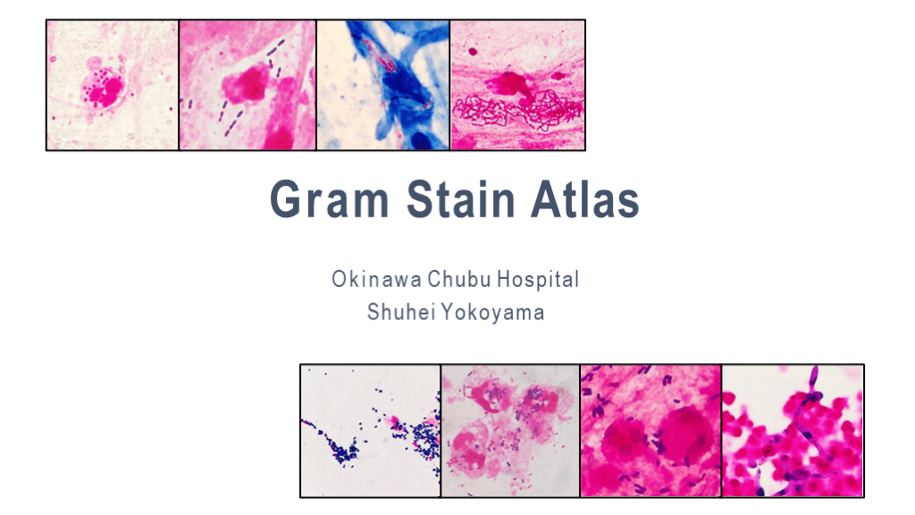
#1. Gram Stain Atlas Okinawa Chubu Hospital Shuhei Yokoyama
#2. Introduction At the Okinawa Chubu Hospital, the gram stain plays a pivotal role in the selection process of the appropriate antibiotic. It is an essential part of our routine.
To become proficient at gram staining, it is vital to have viable specimens, a place to perform the stain, an instructor, and most importantly, ample practice.
The following slides are a collection of sputum gram stain samples. These slides provide a visual guide for the identification of bacteria based on their microscopic characteristics.
肺炎の原因となる一般的な細菌
#3. Common organisms that cause pneumonia: Streptococcus pneumoniae Haemophilus influenzae Moraxella catarrhalis Klebsiella pneumoniae Pseudomonas aeruginosa Staphylococcus aureus
#4. Streptococcus pneumoniae 23.2% of community-acquired pneumonia.
GPDC; Gram-positive lancet-shaped diplococci.
Lancet-shaped
Halos are capsular membranes that may be present around the bacillus. The gram stain reveals a transparent zone surrounding the organism.
Haemophilus influenzaeの特徴
#12. Haemophilus influenzae 18.6% of community-acquired pneumonia.
GNCB; Gram-negative coccobacilli.
Small size
It is difficult to distinguish whether they are a coccus or bacillus due to their small size.
Moraxella catarrhalisの識別
#19. Moraxella catarrhalis 6.1% of community-acquired pneumonia.
GNDC; Gram-negative coccobacilli.
Kidney-shaped
Phagocytic image is remarkable.
It is impossible to distinguish it from other species such as Neisseria meningitidis or Acinetobacter spp. by its shape alone.
Klebsiella pneumoniaeの形態
#29. Klebsiella pneumoniae 3.4% of community-acquired pneumonia.
GNR-large(GNlR); Gran negative large rod.
Box-shaped, stubby, blunted circle.
Pink mucoids may be present around the bacillus.
Mucoids can also be seen in P.aeruginosa.
Pseudomonas aeruginosaの特性
#40. Pseudomonas aeruginosa 3.7% of community-acquired pneumonia.
GNR-small(GNsR); gram-negative small rod.
It is thinner than E.coli.
It can be confused with other gram-negative rods due to the presence of pink mucoids that masks its “thin body” characteristic.
Staphylococcus aureusの分類
#48. Staphylococcus aureus 0.6% of community-acquired pneumonia.
GPC-cluster; gram-positive cocci occurring in clusters.
No morphological distinctions can be made between MSSA and MRSA.
多因子性肺炎の診断
#53. Polymicrobial pattern When both gram positive and gram negative bacteria are found in large numbers, it is difficult to determine the dominant causative organism.
Aspiration pneumonia is suspected when both polymorphonuclear leucocytes (PMN) and numerous squamous epithelial (Epi) are seen.
Corynebacterium spp.の稀な肺炎
#59. Corynebacterium spp. Inverted V-shape.
It is a rare cause of pneumonia. If coryne-form bacteria is seen among other organisms, it is unlikely that Corynebacterium is the causative pathogen.
However, if the gram stain reveals the solitary presence of coryne-form bacteria, pneumonia caused by Corynebacterium can be considered.
Mycobacterium tuberculosisの識別
#64. Mycobacterium tuberculosis Acid fast bacilli (AFB)
気管支上皮細胞と扁平上皮細胞の特徴
#72. Bronchial ciliate cell Characterized by fine whiskers.
A specimen with bronchial ciliate cells suggests that it was taken from the lower respiratory tract.
#76. Squamous epithelial cells Streptococci that originate from the oral cavity can be seen adhered to epithelial cells, but they are not the causative organisms of pneumonia.